| Development of a phantoms for Phase Contrast Tomosynthesisntrast imaging |

|

|

|
|
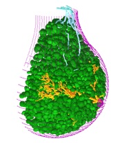
Computational anthropomorphic breast phantom used in phase contrast studies (shown in figure 1).
Figure 1. Mathematical breast phantom
The breast phantom in Figure 1a is a result of a methodology for creation of 3D breast models developed by Bliznakova et al (Bliznakova et al., 2003; Bliznakova et al., 2012; Bliznakova et al., 2010). The methodology consists of algorithms to model the different breast components: (a) external shape; (b) glandular tissue: ducts and Cooper ligaments; (c) adipose tissue; (d) abnormalities like regular and irregular masses, spherical and ellipsoid calcifications and group of micro calcifications (μCas), elongated abnormalities; (e) skin, pectoralis and lymphatics. Briefly described, the breast surface is modeled as a combination of two geometrical primitives: an elongated semi-ellipsoid and an elongated semi-hyperboloid. For breast CT and cone-beam CT applications the external shape may be modeled with a single semi-ellipsoid. The duct system is simulated using a network of cylinders, probabilistically arranged in the breast as branches, in a tree-like arrangement, starting from the nipple and restricted by the external breast contour. The mammographic texture simulates the presence of adipose, fibrous and connective tissues as well as other non-glandular tissue types that are not explicitly modeled. The algorithm is based on the use of random walks, calculated using the concept of the ‘fractional Brownian motion model’, followed by a series of 3D image processing algorithms that result in a realistic 3D texture. Cooper’s ligaments are modeled as a set of thin ellipsoid shells, originating at randomly sampled positions in the breast model, while the pectoralis muscle is approximated as a cone shaped object. Breast abnormalities are modeled with round, ovoid, elongated or irregular shapes. Lymphatic system is included as well. The final breast is a 3D matrix or several 3D matrices composed of voxels with resolution and content defined by the user. The software has been provided to over 30 research and industry organizations and has been regarded as a useful tool to predict the performance of imaging procedures. The package consists of four modules used to create 3D breast models, compress them, simulate their x-ray images and finally visualize the results of the simulations, i.e. 3D breast models and 2D projection images.
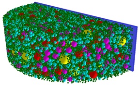
The phantom in Figure 1b simulates an averaged compressed breast and is composed of about 33000 spheres. It represents a software model of the physical phantom developed, validated and tested for 2D mammography and breast tomosynthesis by the LUCMFR group (Lesley et al., 2012). The phantom is composed of two main parts: an acrylic semi-cylinder container of height 58 mm and diameter 200 mm, and equal volumes of acrylic spheres of six different diameters: 15.88, 12.70, 9.52, 6.35, 3.18 and 1.58 mm. As in the physical phantom, the total volume of each different sphere type was set to 60806 mm³.
Bliznakova K, Bliznakov Z, Bravou V, Kolitsi Z and Pallikarakis N 2003 A three-dimensional breast software phantom for mammography simulation Phys Med Biol 48 3699-719 Bliznakova K, Sechopoulos I, Buliev I and Pallikarakis N 2012 BreastSimulator: A software platform for breast x-ray imaging research Journal of Biomedical Graphics and Computing 2 1-14 Bliznakova K, Suryanarayanan S, Karellas A and Pallikarakis N 2010 Evaluation of an improved algorithm for producing realistic 3D breast software phantoms: Application for mammography Medical physics 37 5604-17 Lesley C, Marshall N and and Bosmans H Proceeding IWDM'12 Proceedings of the 11th international conference on Breast Imaging 2012), vol. Series): Springer-Verlag Berlin, Heidelberg ©2012) pp 642-9
|
|||||